our services
Experience board-certified image reviews offering practical and detailed reports. Interpret Radiographs, Ultrasound, Computed Tomography, MRI, and Fluoroscopy. Always accessible through phone or email for consultation or review. A personalized approach that provides a comprehensive and efficient experience for clinicians.
Ultrasonography
Medical Ultrasonography uses ultrasound (high-frequency sound waves) to visualize structures in the body in real time. It is best used for soft tissue structures such as seen in the abdominal cavity or tendons, muscles. No ionizing radiation is involved, but the quality of the images obtained using ultrasound is highly dependent on the skill of the person (ultrasonographer) performing the exam. Ultrasound is limited by its inability to image through air (lungs, bowel loops) or bone with only superficial evaluation available.
Computed Tomography (CT)
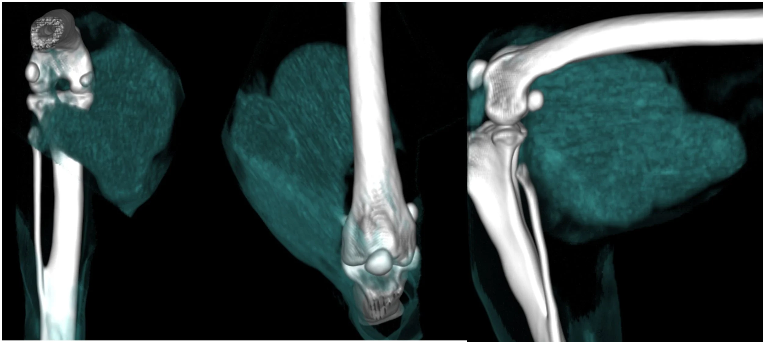
Computed tomography (CT) is a radiologic modality that utilizes ionizing radiation to obtain cross-sectional images of a patient. The images are acquired in the patient’s axial plane (like a loaf of bread) and may also be reprocessed to produce images in many additional anatomic planes or may produce 3-D evaluation of the entire region of interest or individual components such as bone or lungs. Radiocontrast agents are often used with CT for enhanced delineation of anatomy and angiography.
Magnetic Resonance Imaging (MRI)
Magnetic resonance imaging (MRI) is a medical imaging technique that uses a magnetic field and computer-generated radio waves to create detailed images of the organs and tissues in your body. Like CT, MRI is considered cross-sectional imaging and can thus produce 3D images that can be viewed from different angles. MRI in veterinary medicine is primarily used by neurologists and occasionally by surgeons for orthopedic evaluations.
Fluoroscopy
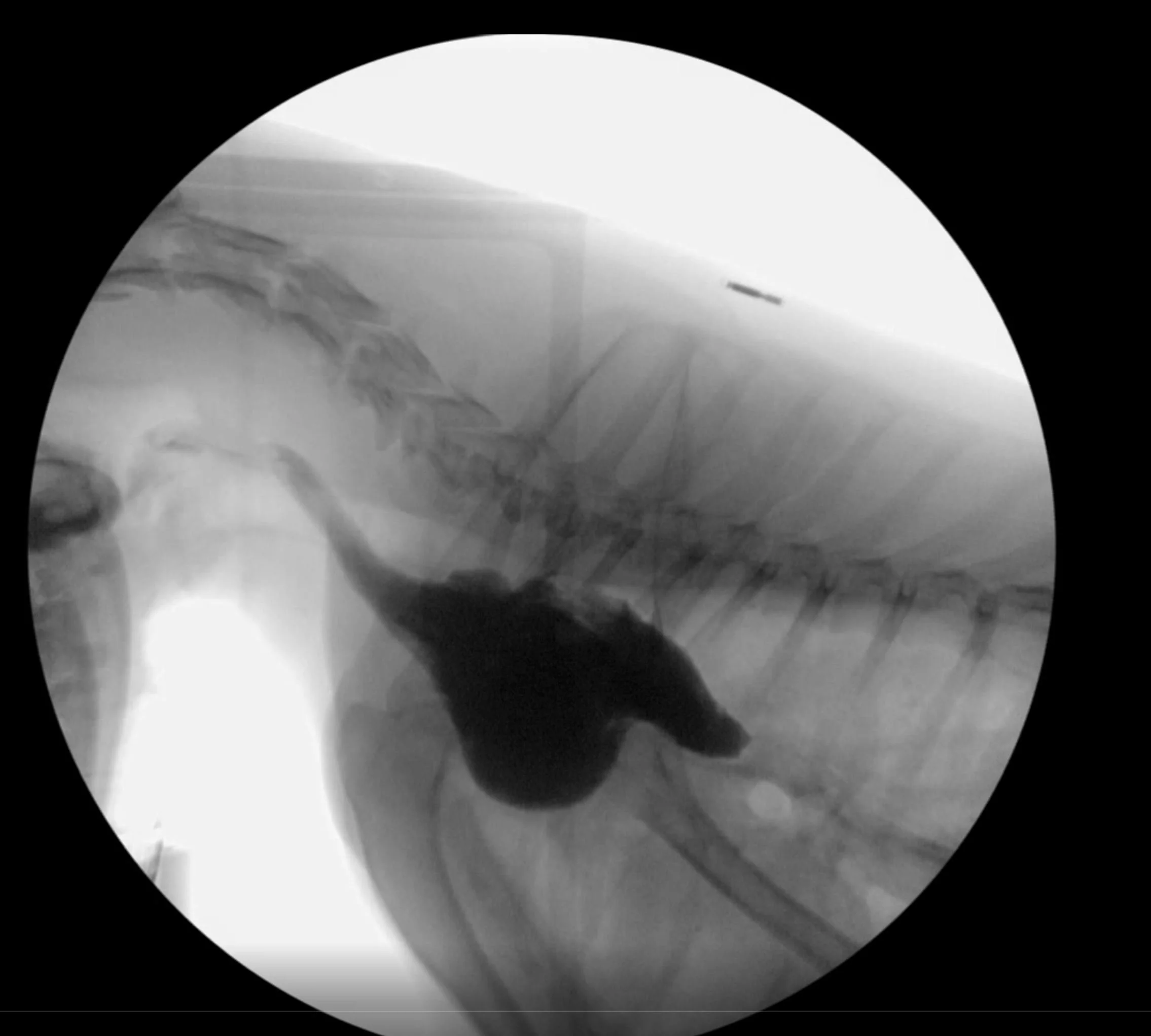
Fluoroscopy is a unique form of imaging that uses a pulses of x-rays to obtain an image but instead of a single image, it produces an x-ray movie or video, in realtime. In veterinary medicine it is used in two primary ways: Treatment such as in orthopaedic surgery for implant placement and second as a diagnostic tool. The latter may include swallowing study, cardiac evaluation, urinary system study to name a few.
Radiographs
Radiographs involve the use of x-rays (ionizing radiation) to produce a two-dimensional image. It is the oldest form of imaging in veterinary medicine and still the most widely used today. Evaluation of both hard and soft tissues is achieved and with proper technique, can provide good quality diagnostic evaluation of the patient.
There is significant variability, however, in the quality in all imaging modalities performed at different sites and by different individuals. Achieving the full potential of all these modalitiesrelies on the level of training of staff and supervising radiologists. Ultimately the quality of the radiographic interpretation is dependent on quality of the images to interpret.


